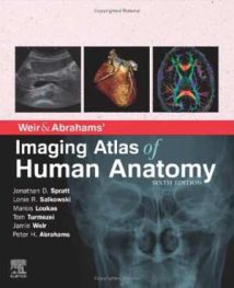
نام کتاب: Weir & Abrahams’ Imaging Atlas Of Human Anatomy
نویسنده: Jonathan D. Spratt و Lonie R Salkowski و Marios Loukas و Tom Turmezei و Jamie Weir و Peter H Abrahams
ویرایش: ۶
سال انتشار: ۲۰۲۰
کد ISBN کتاب: ۹۷۸۰۷۰۲۰۷۹۲۶۹, ۹۷۸۰۷۲۳۴۳۸۲۳۶,
فرمت: PDF
تعداد صفحه: ۲۷۲
حجم کتاب: ۱۴۰ مگابایت
کیفیت کتاب: OCR
انتشارات: Elsevier
Description About Book Weir & Abrahams’ Imaging Atlas Of Human Anatomy From Amazon
Imaging is ever more integral to anatomy education and throughout modern medicine. Building on the success of previous editions, this fully revised fifth edition provides a superb foundation for understanding applied human anatomy, offering a complete view of the structures and relationships within the body using the very latest imaging techniques.
It is ideally suited to the needs of medical students, as well as radiologists, radiographers and surgeons in training. It will also prove invaluable to the range of other students and professionals who require a clear, accurate, view of anatomy in current practice.
Fully revised legends and labels and over 80% new images – featuring the latest imaging techniques and modalities as seen in clinical practice
Covers the full variety of relevant modern imaging – including cross-sectional views in CT and MRI, angiography, ultrasound, fetal anatomy, plain film anatomy, nuclear medicine imaging and more – with better resolution to ensure the clearest anatomical views
Unique new summaries of the most common, clinically important anatomical variants for each body region – reflects the fact that around 20% of human bodies have at least one clinically significant variant
New orientation drawings – to help you understand the different views and the 3D anatomy of 2D images, as well as the conventions between cross-sectional modalities
Now a more compete learning package than ever before, with superb new BONUS electronic enhancements embedded within the accompanying eBook, including:
Labelled image ‘stacks’ – that allow you to review cross-sectional imaging as if using an imaging workstation
Labelled image ‘slide-lines’ – showing features in a full range of body radiographs to enhance understanding of anatomy in this essential modality
Self-test image ‘slideshows’ with multi-tier labelling – to aid learning and cater for beginner to more advanced experience levels
Labelled ultrasound videos – bring images to life, reflecting this increasingly clinically practiced technique
Questions and answers accompany each chapter – to test your understanding and aid exam preparation
۳۴ pathology tutorials – based around nine key concepts and illustrated with hundreds of additional pathology images, to further develop your memory of anatomical structures and lead you through the essential relationships between normal and abnormal anatomy
درباره کتاب Weir & Abrahams’ Imaging Atlas Of Human Anatomy ترجمه شده از گوگل
تصویربرداری همیشه بیشتر در آموزش آناتومی و در کل طب مدرن نقش اساسی دارد. با توجه به موفقیت در چاپ های قبلی ، این نسخه پنجم که کاملاً تجدید نظر شده است ، بنیادی عالی برای درک آناتومی انسانی به کار رفته ارائه می دهد ، و با استفاده از جدیدترین تکنیک های تصویربرداری ، نمای کاملی از ساختارها و روابط درون بدن را ارائه می دهد.
به طور ایده آل متناسب با نیاز دانشجویان پزشکی و همچنین رادیولوژیست ها ، رادیوگرافی ها و جراحان در آموزش است. همچنین برای طیف وسیعی از دانشجویان و متخصصانی که نیاز به دید واضح ، دقیق ، از آناتومی در عمل فعلی دارند ، بسیار ارزشمند خواهد بود.
به طور کامل در افسانه ها و برچسب ها و بیش از ۸۰٪ تصاویر جدید تجدید نظر شده است – دارای جدیدترین تکنیک ها و روش های تصویربرداری همانطور که در عمل بالینی مشاهده می شود
انواع مختلف تصویربرداری مدرن مرتبط را شامل می شود – از جمله نماهای مقطعی در CT و MRI ، آنژیوگرافی ، سونوگرافی ، آناتومی جنین ، آناتومی فیلم ساده ، تصویربرداری پزشکی هسته ای و موارد دیگر – با وضوح بهتر برای اطمینان از واضح ترین دیدگاه های آناتومیک
خلاصه های منحصر به فرد جدید از متداول ترین ، مهمترین انواع آناتومیکی بالینی برای هر منطقه از بدن – این واقعیت را نشان می دهد که حدود ۲۰٪ از بدن انسانها حداقل یک نوع بالینی قابل توجه دارند
نقشه های جدید جهت یابی – برای کمک به شما در درک دیدگاه های مختلف و آناتومی سه بعدی تصاویر ۲D و همچنین قراردادهای بین روش های مقطعی
اکنون یک بسته یادگیری رقابت پذیرتر از هر زمان دیگری ، با پیشرفت های الکترونیکی فوق العاده جدید BONUS که در کتاب الکترونیکی همراه تعبیه شده است ، از جمله:
برچسب «پشته» تصویر – به شما امکان می دهد تصویربرداری مقطعی را مانند استفاده از ایستگاه کاری تصویربرداری مرور کنید
تصاویر دارای برچسب “خطوط اسلاید” – ویژگی هایی را در طیف گسترده ای از رادیوگرافی بدن برای افزایش درک آناتومی در این روش اساسی نشان می دهد
نمایش اسلایدی تصویر خودآزمایی با برچسب گذاری چند لایه – برای کمک به یادگیری و تهیه کردن برای تجربه های مبتدی تا پیشرفته تر
فیلم های سونوگرافی دارای برچسب – تصاویر را زنده می کنند ، و این روش به طور فزاینده ای است که از نظر بالینی انجام می شود
سوالات و پاسخ ها هر فصل را همراهی می کنند – برای آزمایش درک و کمک به آمادگی آزمون
۳۴ آموزش پاتولوژی – مبتنی بر حدود ۹ مفهوم اصلی و نشان داده شده با صدها تصویر آسیب شناسی اضافی ، برای توسعه بیشتر حافظه خود از ساختارهای آناتومیک و هدایت شما از طریق روابط اساسی بین آناتومی طبیعی و غیر طبیعی
[box type=”info”]![]() جهت دسترسی به توضیحات این کتاب در Amazon اینجا کلیک کنید.
جهت دسترسی به توضیحات این کتاب در Amazon اینجا کلیک کنید.![]() در صورت خراب بودن لینک کتاب، در قسمت نظرات همین مطلب گزارش دهید.
در صورت خراب بودن لینک کتاب، در قسمت نظرات همین مطلب گزارش دهید.

