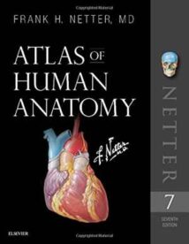
نام کتاب: Atlas Of Human Anatomy
نویسنده: Frank H. Netter
ویرایش: ۷
سال انتشار: ۲۰۱۸
کد ISBN کتاب: ۰۳۲۳۳۹۳۲۲۵, ۹۷۸۰۳۲۳۳۹۳۲۲۵,
فرمت: PDF
تعداد صفحه: ۶۷۲
حجم کتاب: ۱۱۳ مگابایت
کیفیت کتاب: OCR
انتشارات: Elsevier
Description About Book Atlas Of Human Anatomy From Amazon
The only anatomy atlas illustrated by physicians, Atlas of Human Anatomy, 7th edition, brings you world-renowned, exquisitely clear views of the human body with a clinical perspective. In addition to the famous work of Dr. Frank Netter, you’ll also find nearly 100 paintings by Dr. Carlos A. G. Machado, one of today’s foremost medical illustrators. Together, these two uniquely talented physician-artists highlight the most clinically relevant views of the human body. In addition, more than 50 carefully selected radiologic images help bridge illustrated anatomy to living anatomy as seen in everyday practice.
Region-by-region coverage, including Muscle Table appendices at the end of each section.
Large, clear illustrations with comprehensive labels not only of major structures, but also of those with important relationships. Tabular material in separate pages and additional supporting material as a part of the electronic companion so the printed page stays focused on the illustration.
Updates to the 7th ویرایش– based on requests from students and practitioners alike:
New Systems Overview section featuring brand-new, full-body views of surface anatomy, vessels, nerves, and lymphatics.
More than 25 new illustrations by Dr. Machado, including the clinically important fascial columns of the neck, deep veins of the leg, hip bursae, and vasculature of the prostate; and difficult-to-visualize areas like the infratemporal fossa.
New Clinical Tables at the end of each regional section that focus on structures with high clinical significance. These tables provide quick summaries, organized by body system, and indicate where to best view key structures in the illustrated plates.
More than 50 new radiologic images – some completely new views and others using newer imaging tools – have been included based on their ability to assist readers in grasping key elements of gross anatomy.
Updated terminology based on the international anatomic standard, Terminologia Anatomica, with common clinical eponyms included.
Student Consult access includes a pincode to unlock the complete enhanced eBook of the Atlas through Student Consult. Every plate in the Atlas―and over 100 Bonus Plates including illustrations from previous editions―are enhanced with an interactive label quiz option and supplemented with “Plate Pearls” that provide quick key points and supplemental tools for learning, reviewing, and assessing your knowledge of the major themes of each plate. Tools include 300 multiple choice questions, videos, 3D models, and links to related plates.
درباره کتاب Atlas Of Human Anatomy ترجمه شده از گوگل
Atlas of Human Anatomy ، نسخه هفتم ، تنها اطلس آناتومی که توسط پزشکان نشان داده شده است ، دیدگاههای کاملاً مشهور و مشهوری از بدن انسان را با دیدگاه بالینی برای شما به ارمغان می آورد. علاوه بر کار معروف دکتر فرانک نتر ، تقریباً ۱۰۰ نقاشی از دکتر کارلوس ا. ج. ماچادو ، یکی از برجسته ترین تصویرگران پزشکی امروز را نیز خواهید یافت. این دو پزشک-هنرمند با استعداد منحصر به فرد ، در کنار هم مهمترین نظرات مربوط به بدن انسان را برجسته می کنند. علاوه بر این ، بیش از ۵۰ تصویر رادیولوژیک که با دقت انتخاب شده اند ، کمک می کند تا آناتومی مصنوعی را به آناتومی زنده متصل کنید ، همانطور که در عمل روزمره دیده می شود.
پوشش منطقه به منطقه ، از جمله پیوست های جدول عضله در انتهای هر بخش.
تصاویر بزرگ و واضح با برچسب های جامع نه تنها از ساختارهای اصلی ، بلکه از کسانی که روابط مهمی نیز دارند. مواد جداولی در صفحات جداگانه و مواد پشتیبانی اضافی به عنوان بخشی از همراه الکترونیکی ، بنابراین صفحه چاپ شده روی تصویر متمرکز است.
به روزرسانی های نسخه ۷ – براساس درخواست دانشجویان و پزشکان به طور یکسان:
بخش نمای کلی سیستم های جدید شامل نمای کاملاً جدید و تمام بدن از آناتومی سطح ، عروق ، اعصاب و لنفاوی.
بیش از ۲۵ تصویر جدید توسط دکتر ماچادو ، از جمله ستون های مهم فاسیال گردن ، رگهای عمیق پا ، سوراخ های مفصل ران و عروق پروستات. و مناطقی که به سختی قابل تجسم هستند مانند حفره زیر زمان.
جداول بالینی جدید در پایان هر بخش منطقه ای که بر ساختارهایی با اهمیت بالینی بالا تمرکز دارند. این جداول خلاصه های سریعی را که توسط سیستم بدنی سازمان یافته اند ، ارائه می دهند و نشان می دهند که کجا ساختارهای اصلی را در صفحه های نشان داده شده به بهترین شکل مشاهده کنید.
بیش از ۵۰ تصویر رادیولوژی جدید – برخی از نماهای کاملاً جدید و برخی دیگر با استفاده از ابزارهای تصویربرداری جدیدتر – بر اساس توانایی آنها در کمک به خوانندگان در درک عناصر اصلی آناتومی ناخالص گنجانده شده است.
اصطلاحات به روز شده بر اساس استاندارد آناتومیک بین المللی ، Terminologia Anatomica ، با نام مستعار بالینی مشترک.
دسترسی Student Consult شامل یک کد رمز برای باز کردن کامل کتاب الکترونیکی پیشرفته اطلس از طریق Student Student است. هر صفحه در اطلس ― و بیش از ۱۰۰ صفحه پاداش شامل تصاویر از نسخه های قبلی ― با یک گزینه مسابقه برچسب تعاملی افزایش یافته و با “مروارید صفحه” که نکات کلیدی سریع و ابزارهای مکمل برای یادگیری ، بررسی و ارزیابی دانش شما از موضوعات اصلی هر صفحه. ابزارها شامل ۳۰۰ س questionsال چند گزینه ای ، فیلم ، مدل سه بعدی و پیوند به صفحات مرتبط هستند.
[box type=”info”]![]() جهت دسترسی به توضیحات این کتاب در Amazon اینجا کلیک کنید.
جهت دسترسی به توضیحات این کتاب در Amazon اینجا کلیک کنید.![]() در صورت خراب بودن لینک کتاب، در قسمت نظرات همین مطلب گزارش دهید.
در صورت خراب بودن لینک کتاب، در قسمت نظرات همین مطلب گزارش دهید.

