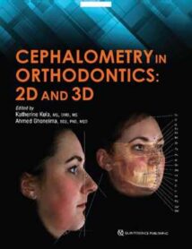
نام کتاب: Cephalometry In Orthodontics – 2D And 3D
نویسنده: Adjunct Facility و Katherine Kula و Ph.D. Ghoneima و Ahmed
ویرایش: ۱
سال انتشار: ۲۰۱۸
کد ISBN کتاب: ۰۸۶۷۱۵۷۶۲۳, ۹۷۸۰۸۶۷۱۵۷۶۲۸,
فرمت: PDF
تعداد صفحه: ۲۰۲
حجم کتاب: ۱۰۱ مگابایت
کیفیت کتاب: OCR
انتشارات: Quintessence Pub Co
Description About Book Cephalometry In Orthodontics – 2D And 3D From Amazon
Cephalometrics has been used for decades to diagnose orthodontic problems and evaluate treatment. However, the shift from 2D to 3D radiography has left some orthodontists unsure about how to use this method effectively. This book defines and depicts all cephalometric landmarks on a skull or spine in both 2D and 3D and then identifies them on radiographs. Each major cephalometric analysis is described in detail, and the linear or angular measures are shown pictorially for better understanding. Because many orthodontists pick specific measures from various cephalometric analyses to formulate their own analysis, these measures are organized relative to the skeletal or dental structure and then compared or contrasted relative to diagnosis, growth, and treatment. Cephalometric norms (eg, age, sex, ethnicity) are also discussed relative to treatment and esthetics. The final chapter shows the application of these measures to clinical cases to teach clinicians and students how to use them effectively. As radiology transitions from 2D to 3D, it is important to evaluate the efficacy and cost-effectiveness of each in diagnosis and treatment, and this book outlines all of the relevant concerns for daily practice.Contents 1. Introduction to the Use of Cephalometrics 2. 2D and 3D Radiography 3. Skeletal Landmarks and Measures 4. Frontal Cephalometric Analysis 5. Soft Tissue Analysis 6. A Perspective on Norms and Standards 7. The Transition from 2D to 3D Cephalometrics: Understanding the Problems of Landmarks and Measures 8. Cephalometric Airway Analysis 9. Radiographic Superimposition: From 2D to 3D 10. Growth and Treatment Predictions: Accuracy and Reliability 11. Measuring Bone with CBCT 12. Common Pathologic Findings in Cephalometric Radiology 13. The Cost of 2D Versus 3D Radiology 14. Clinical Cases
درباره کتاب Cephalometry In Orthodontics – 2D And 3D ترجمه شده از گوگل
سفالومتریک برای دهه ها برای تشخیص مشکلات ارتودنسی و ارزیابی درمان مورد استفاده قرار گرفته است. با این حال ، تغییر از رادیوگرافی ۲D به ۳D باعث شده برخی از ارتودنتیست ها در مورد چگونگی استفاده موثر از این روش مطمئن نباشند. این کتاب کلیه نشانه های سفالومتریک روی جمجمه یا ستون فقرات را به صورت ۲D و ۳D تعریف کرده و به تصویر می کشد و سپس آنها را در رادیوگرافی شناسایی می کند. هر تجزیه و تحلیل سفالومتریک عمده با جزئیات شرح داده شده است ، و اندازه گیری های خطی یا زاویه ای برای درک بهتر به صورت تصویری نشان داده شده است. از آنجا که بسیاری از متخصصان ارتودنسی اقدامات ویژه ای را از روش های مختلف تجزیه و تحلیل سفالومتری برای تهیه تجزیه و تحلیل خود انجام می دهند ، این اقدامات نسبت به ساختار اسکلتی یا دندانی سازمان یافته و سپس نسبت به تشخیص ، رشد و درمان با یکدیگر مقایسه می شوند. هنجارهای سفالومتری (به عنوان مثال ، سن ، جنس ، قومیت) نیز در رابطه با درمان و زیبایی مورد بحث قرار می گیرند. فصل آخر کاربرد این اقدامات را در موارد بالینی برای آموزش پزشکان و دانشجویان به نحوه استفاده موثر از آنها نشان می دهد. همانطور که رادیولوژی از ۲D به ۳D منتقل می شود ، ارزیابی اثربخشی و مقرون به صرفه بودن هر یک در تشخیص و درمان مهم است ، و این کتاب تمام نگرانی های مربوط به تمرین روزانه را بیان می کند. مطالب ۱٫ مقدمه ای در استفاده از سفالومتریک ۲٫ رادیوگرافی ۲D و ۳D 3. شاخص های اسکلتی و اقدامات ۴٫ تجزیه و تحلیل سفالومتری فرونتال ۵٫ تجزیه و تحلیل بافت نرم ۶٫ دیدگاه در مورد هنجارها و استانداردها ۷٫ انتقال از سفالومتری ۲D به ۳D: درک مشکلات نشانه ها و اقدامات ۸٫ تجزیه و تحلیل راه هوایی سفالومتریک ۹٫ سوپرمیشن رادیوگرافی: از ۲D به ۳ بعدی ۱۰٫ رشد و درمان پیش بینی ها: دقت و اطمینان ۱۱٫ اندازه گیری استخوان با CBCT 12. یافته های پاتولوژیک رایج در رادیولوژی سفالومتریک ۱۳٫ هزینه ۲D در مقابل رادیولوژی ۳D 14. موارد بالینی
[box type=”info”]![]() جهت دسترسی به توضیحات این کتاب در Amazon اینجا کلیک کنید.
جهت دسترسی به توضیحات این کتاب در Amazon اینجا کلیک کنید.![]() در صورت خراب بودن لینک کتاب، در قسمت نظرات همین مطلب گزارش دهید.
در صورت خراب بودن لینک کتاب، در قسمت نظرات همین مطلب گزارش دهید.

