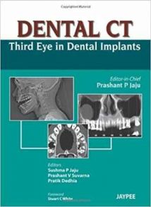
نام کتاب: Dental Ct – Third Eye In Dental Implants
نویسنده: Pratik Dedhia و Prashant P. Jaju و Sushma P. Jaju و Prashant V. Suvarna
ویرایش: ۱
سال انتشار: ۲۰۱۳
کد ISBN کتاب: ۹۷۸۹۳۵۱۵۲۰۲۴۵, ۹۳۵۱۵۲۰۲۴۲,
فرمت: PDF
تعداد صفحه: ۷۰
حجم کتاب: ۱۵ مگابایت
کیفیت کتاب: OCR
انتشارات: Jaypee Brothers
Description About Book Dental Ct – Third Eye In Dental Implants From Amazon
Dental implants are the future of dentistry. Oral radiology is the third dimension for a successful dental implant practice. Two-dimensional imaging shares its limitations in dental implantology which was easily overcome by the advent of dental CT. The 3-D imaging provides a clear relationship between structures that could be obscure on 2-D images. CBCT is useful for assessing impacted teeth, particularly the relationship between mandibular third molars and mandibular canals. It is also valuable in assessing implant positioning and preimplant bone augmentation to provide the best possible prosthodontics reconstructive outcome. This book is particularly useful for demonstrating the value of 3-D imaging for the specific purpose of dental implant planning. Describes the various dental programs optimized for dental applications of computed tomography, in particular focuses on the utility of dental CT for implantology oral and maxillofacial surgery, endodontics, periodontics, advances in implant imaging and case studies. Emphasises on basic principles and detailed description of the steps for each examination methods such as how to position the patient and interpret the images for getting desired result. Qualities of the images are high resolution including both normal anatomic structures in the regions of interest and various common pathologic conditions. The principles and examples of radiographic interpretation are fully applicable to cone-beam imaging. This book is aimed both at “old dogs” and “new dogs” to dental CT in implantology.
درباره کتاب Dental Ct – Third Eye In Dental Implants ترجمه شده از گوگل
کاشت دندان آینده دندانپزشکی است. رادیولوژی دهان بعد سوم برای موفقیت آمیز بودن عمل کاشت دندان است. تصویربرداری دو بعدی محدودیت های خود را در ایمپلنتولوژی دندان دارد که با ظهور CT دندان به راحتی برطرف شد. تصویربرداری ۳-D یک رابطه واضح بین ساختارهایی است که می تواند در تصاویر دو بعدی مبهم باشد. CBCT برای ارزیابی دندانهای نهفته ، به ویژه رابطه بین دندانهای مولر سوم فک پایین و کانالهای فک پایین مفید است. همچنین در ارزیابی موقعیت ایمپلنت و افزایش استخوان قبل از لانه گزینی برای ارائه بهترین نتیجه ترمیمی پروتزهای دندانی بسیار ارزشمند است. این کتاب به ویژه برای نشان دادن ارزش تصویربرداری ۳-D برای هدف خاص برنامه ریزی ایمپلنت دندان بسیار مفید است. برنامه های مختلف دندانپزشکی را که برای کاربردهای دندانپزشکی توموگرافی کامپیوتری بهینه شده است ، توصیف می کند ، به ویژه بر استفاده از CT دندان برای جراحی دهان و فک و صورت ، ریشه دندان ، پریودنتزی ، پیشرفت در تصویربرداری ایمپلنت و مطالعات موردی تمرکز دارد. بر اصول اساسی و شرح دقیق مراحل هر روش معاینه مانند نحوه قرار دادن بیمار و تفسیر تصاویر برای گرفتن نتیجه مطلوب تأکید دارد. کیفیت کتابتصاویر با وضوح بالا شامل ساختارهای آناتومیک طبیعی در مناطق مورد نظر و شرایط مختلف پاتولوژیک مختلف است. اصول و نمونه های تفسیر رادیوگرافی به طور کامل در تصویربرداری پرتو مخروطی قابل استفاده است. این کتاب با هدف “سگهای پیر” و “سگهای جدید” برای CT دندان در ایمپلنتولوژی انجام شده است.
[box type=”info”]![]() جهت دسترسی به توضیحات این کتاب در Amazon اینجا کلیک کنید.
جهت دسترسی به توضیحات این کتاب در Amazon اینجا کلیک کنید.![]() در صورت خراب بودن لینک کتاب، در قسمت نظرات همین مطلب گزارش دهید.
در صورت خراب بودن لینک کتاب، در قسمت نظرات همین مطلب گزارش دهید.

