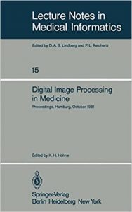
نام کتاب: Digital Image Processing In Medicine
نویسنده: Prof. Dr. P. H. Heintzen و R. Brennecke و Karl Heinz Höhne
ویرایش: ۱
سال انتشار: ۱۹۸۱
کد ISBN کتاب: ۹۷۸۳۵۴۰۱۰۸۷۷۱, ۹۷۸۳۶۴۲۹۳۱۸۸۸
فرمت: PDF
تعداد صفحه: ۱۹۷
انتشارات: Springer-Verlag Berlin Heidelberg
Description About Book Digital Image Processing In Medicine From Amazon
In diagnostic medicine a large part of information about the patient is drawn from data, which, more or less, are represented in an opti calor pictorial form. There is a very wide range of such data as e.g. the patients appearance, the various kinds of radiological images, or cytological imagery. In conventional diagnostics the data, as it comes from the acquisition device, is perceived by the physician and is interpreted with the help of a large amount of “a priori” knowledge to give a diagnostic finding. During the last 15 years a steadily rising number of attempts have been made to support these processes by the application of com puters. The attempts mainly concentrate on three objectives: 1. Support of the perception process by the production of better or new types of images, e.g. by Computer tomography or Computer angio graphy (image processing) . 2. Automation of the interpretation process, e.g. for bloodcell dif ferentiation (pattern recognition) . 3. Management of the steeply rising amount of medical image data in the hospital (image data bases) . Although the early applications of digital methods aimed at the second . . objective, in the last years much more success has been a achieved in the support of the perception process by methods of image process ing. The reason for this is obvious – in the case of automatic interpre tation the a priori knowledge of the physician has to be formalized.
درباره کتاب Digital Image Processing In Medicine ترجمه شده از گوگل
در دارو های تشخیصی بخش بزرگی از اطلاعات در مورد بیمار از داده ها، که، بیشتر یا کمتر، در یک CALOR شکل تصویری OPTI نشان کشیده شده است. است محدوده بسیار گسترده ای از داده ها مانند به عنوان مثال، وجود دارد ظاهر بیماران، انواع مختلف تصاویر رادیولوژی، و یا تصاویر سیتولوژی. در تشخیص متعارف داده ها، به عنوان آن را از دستگاه جمع آوری می آید، توسط پزشک درک شده و با کمک یک مقدار زیادی از “پیشینی” دانش تفسیر به یک یافته تشخیصی. در طول ۱۵ سال گذشته تعداد به طور پیوسته رو به افزایش تلاش شده برای حمایت از این فرآیندهای با استفاده از puters کام. تلاش به طور عمده در سه هدف تمرکز: ۱٫ پشتیبانی از فرایند ادراک را با تولید انواع بهتر یا جدید از تصاویر، به عنوان مثال، توسط توموگرافی کامپیوتری یا گرافی کامپیوتر آنژیو (پردازش تصویر). ۲٫ اتوماسیون فرایند تفسیر، به عنوان مثال، برای ferentiation تومان bloodcell (تشخیص الگو). ۳٫ مدیریت از مقدار شدت رو به افزایش از داده های تصویر پزشکی در بیمارستان (پایگاه داده های تصویر). اگر چه برنامه های اولیه از روش دیجیتال با هدف دوم. . هدف، در سال گذشته موفقیت خیلی بیشتر است به دست آمده در حمایت از فرایند ادراک را با استفاده از روش پردازش تصویر ING بوده است. دلیل این کار این واضح است – در مورد tation interpre خودکار دانش پیشینی پزشک تا به رسمیت.
[box type=”info”]![]() جهت دسترسی به توضیحات این کتاب در Amazon اینجا کلیک کنید.
جهت دسترسی به توضیحات این کتاب در Amazon اینجا کلیک کنید.![]() در صورت خراب بودن لینک کتاب، در قسمت نظرات همین مطلب گزارش دهید.
در صورت خراب بودن لینک کتاب، در قسمت نظرات همین مطلب گزارش دهید.

