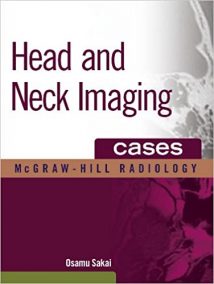
نام کتاب: Head And Neck Imaging Cases
نویسنده: Osamu Sakai
ویرایش: ۱
سال انتشار: ۲۰۱۱
کد ISBN کتاب: ۰۰۷۱۷۸۵۰۲۷, ۹۷۸۰۰۷۱۷۸۵۰۲۰
فرمت: PDF
تعداد صفحه: ۱۲۷۲
انتشارات: Mcgraw-Hill Education / Medical
Description About Book Head And Neck Imaging Cases From Amazon
۳۶۶ cases and more than 3000 images help you accurately interpret head and neck imaging
Head and Neck Imaging Cases uses 366 cases and more than 3000 images to familiarize you with imaging findings of common head and neck diseases and conditions encountered in daily practice. Rarer diseases that have typical image findings as well as normal variants and benign conditions that may be mistaken as abnormalities or malignancies are also included. Reflecting real-world practice, CT and MRI are the main modalities illustrated throughout the book. In addition, you will find cases utilizing fluoroscopy, PET-CT, conventional angiogram/interventional radiology, and radiotherapy/radiosurgery.
The book’s easy-to-navigate organization is specifically designed for use at the workstation. The concise, quick-scan text, numerous images, helpful icons, and pearls speed and simplify the learning process.
Features:
Cases involve the temporal bones, skull base, nasal cavity, and paranasal sinuses, orbit, globe, suprahyoid neck, salivary gland, oral cavity and oropharynx, jaw, larynx and hypopharynx, infrahyoid neck, and lymph nodes Each case includes presentation, findings, differential diagnosis, boxed pearls, and numerous images Icons, a grading system depicting the full spectrum of findings from common to rare and typical to unusual along with consistent chapter organization make this perfect for rapid at-the-bench consultation
درباره کتاب Head And Neck Imaging Cases ترجمه شده از گوگل
۳۶۶ نفر و بیش از ۳۰۰۰ تصاویر به شما کمک کند با دقت تفسیر تصویربرداری سر و گردن
سر و گردن تصویربرداری مخازن با استفاده از ۳۶۶ نفر و بیش از ۳۰۰۰ تصاویر شما را آشنا با تصویربرداری یافته های سر بیماریهای مشترک گردن و شرایط مواجه می شوند در عمل روزانه. بیماری های نادر است که یافته های تصویر معمولی و همچنین انواع نرمال و خوش خیم است که ممکن است اشتباه به عنوان ناهنجاری و یا بدخیمی ها نیز گنجانده شده است. بازتاب عمل در دنیای واقعی، CT و MRI روش اصلی نشان داده شده در سراسر این کتاب می باشد. علاوه بر این، شما موارد استفاده از فلوروسکوپی، PET-CT، آنژیوگرام معمولی / رادیولوژی مداخله ای، و پرتودرمانی / پرتو جراحی پیدا
سازمان آسان به حرکت به کتاب است به طور خاص برای استفاده در ایستگاه های کاری طراحی شده است. مختصر، متن سریع اسکن، تصاویر متعدد، آیکون های مفید، و سرعت مروارید و ساده فرایند یادگیری است.
امکانات:
موارد شامل استخوان گیجگاهی، قاعده جمجمه، حفره بینی، و پارانازال سینوس ها، مدار، جهان، گردن suprahyoid، غدد بزاقی، حفره دهان و اوروفارنکس، فک، حنجره و هیپوفارنکس، گردن infrahyoid، و غدد لنفاوی هر مورد شامل ارائه، یافته ها، تشخیص های افتراقی، مروارید بسته بندی، و تصاویر متعدد آیکن ها، یک سیستم درجه بندی به تصویر می کشد که طیف کاملی از یافته های مشترک برای نادر و معمولی به غیر معمول همراه با سازمان فصل سازگار این مناسب برای سریع مشاوره در از حد نیمکت
[box type=”info”]![]() جهت دسترسی به توضیحات این کتاب در Amazon اینجا کلیک کنید.
جهت دسترسی به توضیحات این کتاب در Amazon اینجا کلیک کنید.![]() در صورت خراب بودن لینک کتاب، در قسمت نظرات همین مطلب گزارش دهید.
در صورت خراب بودن لینک کتاب، در قسمت نظرات همین مطلب گزارش دهید.

