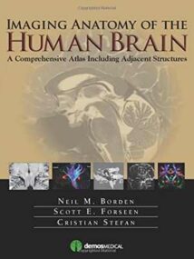
نام کتاب: Imaging Anatomy Of The Human Brain – A Comprehensive Atlas Including Adjacent Structures
نویسنده: Neil M. Borden و Scott E. Forseen و Cristian Stefan
ویرایش: ۱
سال انتشار: ۲۰۱۵
کد ISBN کتاب: ۱۹۳۶۲۸۷۷۴۹, ۹۷۸۱۹۳۶۲۸۷۷۴۱,
فرمت: PDF
تعداد صفحه: ۴۶۴
حجم کتاب: ۵۰ مگابایت
کیفیت کتاب: OCR
انتشارات: Demos Medical
Description About Book Imaging Anatomy Of The Human Brain – A Comprehensive Atlas Including Adjacent Structures From Amazon
The most precise, cutting-edge images of normal cerebral anatomy available today are the centerpiece of this spectacular atlas for clinicians, trainees, and students in the neurologically-based medical and non-medical specialties. Truly an ìatlas for the 21st century,î this comprehensive visual reference presents a detailed overview of cerebral anatomy acquired through the use of multiple imaging modalities including advanced techniques that allow visualization of structures not possible with conventional MRI or CT. Beautiful color illustrations using 3-D modeling techniques based upon 3D MR volume data sets further enhances understanding of cerebral anatomy and spatial relationships. The anatomy in these color illustrations mirror the black and white anatomic MR images presented in this atlas. Written by two neuroradiologists and an anatomist who are also prominent educators, along with more than a dozen contributors, this state-of-the-art atlas will serve as an authoritative learning tool in the classroom, and as an invaluable practical resource at the workstation or in the office or clinic.
درباره کتاب Imaging Anatomy Of The Human Brain – A Comprehensive Atlas Including Adjacent Structures ترجمه شده از گوگل
دقیق ترین و برترین تصاویر آناتومی مغزی طبیعی که امروزه در دسترس است ، قطب اصلی این اطلس تماشایی برای پزشکان ، کارآموزان و دانشجویان متخصص پزشکی و غیرپزشکی مبتنی بر عصب شناسی است. این مرجع بصری واقعاً “یک اطلس برای قرن بیست و یکم” ، یک نمای کلی از آناتومی مغزی را که از طریق استفاده از چندین روش تصویربرداری از جمله تکنیک های پیشرفته که تجسم ساختارهایی را که با MRI یا CT معمولی امکان پذیر نیست ، به دست می آورد ، ارائه می دهد. تصاویر زیبای رنگی با استفاده از تکنیک های مدل سازی ۳-D بر اساس مجموعه داده های حجم ۳D MR ، درک آناتومی مغزی و روابط فضایی را بیشتر افزایش می دهد. آناتومی در این تصاویر رنگی تصاویر MR آناتومیک سیاه و سفید را در این اطلس منعکس می کند. این اطلس پیشرفته که توسط دو متخصص نورورادیولوژیست و یک آناتومیست که همچنین مربیان برجسته ای هستند ، به همراه بیش از دوازده همکار ، نوشته شده است به عنوان یک ابزار یادگیری معتبر در کلاس ، و به عنوان یک منبع عملی ارزشمند در ایستگاه کاری خدمت می کند یا در مطب یا کلینیک.
[box type=”info”]![]() جهت دسترسی به توضیحات این کتاب در Amazon اینجا کلیک کنید.
جهت دسترسی به توضیحات این کتاب در Amazon اینجا کلیک کنید.![]() در صورت خراب بودن لینک کتاب، در قسمت نظرات همین مطلب گزارش دهید.
در صورت خراب بودن لینک کتاب، در قسمت نظرات همین مطلب گزارش دهید.

