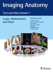
نام کتاب: Imaging Anatomy – Text And Atlas Volume 1, Lungs, Mediastinum, And Heart
نویسنده: Farhood Saremi و Damian Sanchez-Quintana و Hiro Kiyosue و Francesco Faletra و Meng Law و Dakshesh Patel و R. Shane Tubbs
ویرایش: ۱
سال انتشار: ۲۰۲۱
کد ISBN کتاب: ۲۰۲۱۰۰۱۹۱۵, ۲۰۲۱۰۰۱۹۱۶, ۹۷۸۱۶۲۶۲۳۹۸۸۳, ۹۷۸۱۶۲۶۲۳۹۸۹۰,
فرمت: EPUB
تعداد صفحه: ۵۹۸
حجم کتاب: ۸۵ مگابایت
کیفیت کتاب: OCR
انتشارات: Thieme
Description About Book Imaging Anatomy – Text And Atlas Volume 1, Lungs, Mediastinum, And Heart From Amazon
First volume in state-of-the-art radiologic text-atlas series details anatomy of the lungs, mediastinum, and heart
Normal imaging anatomy and variants, including both diagnostic and surgical anatomy, are the cornerstones of radiologic knowledge. Imaging Anatomy: Text and Atlas Volume 1, Lungs, Mediastinum, and Heart is the first in a series of four richly illustrated radiologic references edited by distinguished radiologist Farhood Saremi and coedited by Damian Sanchez-Quintana, Hiro Kiyosue, Francesco F. Faletra, Meng Law, Dakshesh Patel, and Shane Tubbs, with contributions from an impressive cadre of international authors.
The exquisitely crafted atlas provides high-quality multiplanar and volumetric color-coded imaging techniques utilizing CT, MRI, or angiography, supplemented by cadaveric presentations and color drawings that best elucidate each specific anatomic region. Twenty-one chapters with concise text encompass thoracic wall, mediastinum, lung, vascular, and cardiac anatomy, providing readers with a virtual dissection experience. Many anatomical variants along with pathological examples are presented.
Key Highlights
More than 600 illustrations enhance understanding of impacted regions
Lung anatomy including the pleura, pulmonary arteries, pulmonary veins, and lymphatics
Discussion of the tracheobronchial system, mediastinum and thymus, thoracic aorta and major branches, systemic veins, lymphatics and nerves of the thorax, diaphragm, and breast
Heart anatomy including the atrioventricular septal region; aortic, pulmonary, mitral and tricuspid valves; coronary arteries and myocardial perfusion; coronary veins; and pericardium
This superb resource is essential reading for medical students, radiology residents and veteran radiologists, cardiologists, as well as cardiovascular and thoracic surgeons. It provides an excellent desk reference and practical guide for differentiating normal versus pathologic anatomy.
This book includes complimentary access to a digital copy on https://medone.thieme.com.
درباره کتاب Imaging Anatomy – Text And Atlas Volume 1, Lungs, Mediastinum, And Heart ترجمه شده از گوگل
جلد اول در مجموعه متون رادیولوژیکی متون اتلس جزئیات آناتومی ریه ها ، مدیاستینوم و قلب
آناتومی عادی تصویربرداری و انواع آن ، از جمله آناتومی تشخیصی و جراحی ، سنگ بنای دانش رادیولوژی هستند. تصویربرداری آناتومی: متن و اطلس جلد ۱ ، ریه ها ، مدیاستینوم و قلب اولین مورد از سری چهار مرجع پرتوی رادیولوژیکی است که توسط رادیولوژیست فرهاد صارمی ویرایششده و توسط دامیان سانچز-کوئینتانا ، هیرو کیوسوئه ، فرانچسکو فالتره ، منگ ویرایششده است. Law ، Dakshesh Patel و Shane Tubbs ، با مشارکت کادر چشمگیر نویسندهبین المللی.
اطلس فوق العاده ساخته شده ، تکنیک های تصویربرداری چند صفحه ای و حجمی با کیفیت کتاببالا با استفاده از CT ، MRI یا آنژیوگرافی را ارائه می دهد ، که با ارائه جسد و نقاشی های رنگی که هر منطقه آناتومیکی خاص را به بهترین وجه روشن می کند ، تکمیل می شود. بیست و یک فصل با متن مختصر شامل دیواره قفسه سینه ، مدیاستینوم ، ریه ، عروق و آناتومی قلبی است و تجربه تشریح مجازی را در اختیار خوانندگان قرار می دهد. انواع مختلف تشریحی به همراه نمونه های آسیب شناسی ارائه شده است.
نکات برجسته کلیدی
بیش از ۶۰۰ تصویر درک مناطق تحت تأثیر را افزایش می دهد
آناتومی ریه شامل پلور ، شریان های ریوی ، وریدهای ریوی و لنفاوی
بحث در مورد سیستم تراشه برونش ، مدیاستین و تیموس ، آئورت قفسه سینه و شاخه های اصلی ، وریدهای سیستمیک ، لنفاوی و اعصاب قفسه سینه ، دیافراگم و سینه
آناتومی قلب شامل ناحیه تیغه دهلیزی – بطنی ؛ دریچه های آئورت ، ریوی ، میترال و سه قلو ؛ عروق کرونر و پرفیوژن میوکارد ؛ وریدهای کرونری ؛ و پریکارد
این منبع عالی برای دانشجویان پزشکی ، متخصصان رادیولوژی و رادیولوژیست های کهنه ، قلب و همچنین جراحان قلب و عروق و قفسه سینه ضروری است. این یک مرجع روی میز عالی و راهنمای عملی برای تمایز آناتومی طبیعی و پاتولوژیک است.
این کتاب شامل دسترسی رایگان به نسخه دیجیتال در https://medone.thieme.com است.
[box type=”info”]![]() جهت دسترسی به توضیحات این کتاب در Amazon اینجا کلیک کنید.
جهت دسترسی به توضیحات این کتاب در Amazon اینجا کلیک کنید.![]() در صورت خراب بودن لینک کتاب، در قسمت نظرات همین مطلب گزارش دهید.
در صورت خراب بودن لینک کتاب، در قسمت نظرات همین مطلب گزارش دهید.

