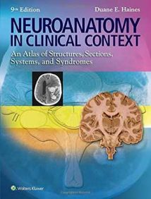
نام کتاب: Neuroanatomy In Clinical Context – An Atlas Of Structures, Sections, Systems, And Syndromes
نویسنده: Duane E. Haines
ویرایش: ۹
سال انتشار: ۲۰۱۴
کد ISBN کتاب: ۱۴۵۱۱۸۶۲۵۸, ۹۷۸۱۴۵۱۱۸۶۲۵۳,
فرمت: PDF
تعداد صفحه: ۳۶۸
حجم کتاب: ۳۱ مگابایت
کیفیت کتاب: OCR
انتشارات: Lww
Description About Book Neuroanatomy In Clinical Context – An Atlas Of Structures, Sections, Systems, And Syndromes From Amazon
Neuroanatomy in Clinical Context, Ninth ویرایشprovides everything the student needs to master the anatomy of the central nervous system, all in a clinical setting. Clear explanations;
abundant MRI, CT, MRA, and MRV images; full-color photographs and illustrations; hundreds of review questions; and supplemental online resources combine to provide a sound anatomical base for integrating neurobiological and clinical concepts. In thus applying neuroanatomy clinically, the atlas ensures student preparedness for exams and for rotations. This authoritative approach—combined with such salutary features as full-color stained sections, extensive cranial nerve cross-referencing, and systems neurobiology coverage—sustains the legacy of this revolutionary teaching and learning tool as the neuroanatomy atlas.
New and hallmark features elucidate neuroanatomy and systems neurobiology for course success!NEW! Chapter on Herniation Syndromes decodes the elegant relationship between brain injury and resulting deficit.NEW! Clinical information integrated throughout the text is screened in blue for quick identification on the page.NEW! Enhanced clinical images emphasize clarity and detail like never before, including full-color images replacing many in black and white, higher-resolution brain scans, and reprocessed spinal cord and brainstem images.MRIs complement full-color anatomical illustrations, allowing for visualization of structures both as they appear to the unaided eye and on imaging studies.Unique, full-color illustrations integrate clinical images of representative lesions with the corresponding deficits highlighted.Full-color stained sections facilitate the easy identification of anatomical features.Dozens of pathway drawings superimposed over MRIs connect structure with function of neural pathways.
Located on thePoint, this atlas’s companion website offers a variety of supplemental
learning resources to maximize study and review time!Question bank featuring over 280 USMLE-style and chapter-review style questionsBonus dissection photographs and brain slice series
درباره کتاب Neuroanatomy In Clinical Context – An Atlas Of Structures, Sections, Systems, And Syndromes ترجمه شده از گوگل
عصب کشی در متن بالینی ، ویرایشنهم ، هر آنچه دانشجو برای تسلط بر آناتومی سیستم عصبی مرکزی نیاز دارد را فراهم می کند ، همه در یک شرایط بالینی. توضیحات روشن
تصاویر فراوان MRI ، CT ، MRA و MRV. عکس ها و تصاویر تمام رنگی ؛ صدها سوال مرور و منابع آنلاین مکمل با هم ترکیب می شوند و یک پایگاه تشریحی صدا برای ادغام مفاهیم عصبی و بالینی فراهم می کنند. در نتیجه اعمال بالینی عصب کشی ، اطلس آمادگی دانش آموزان را برای امتحانات و چرخش ها تضمین می کند. این رویکرد معتبر – همراه با ویژگی های سلامتی مانند بخش های رنگ آمیزی شده تمام رنگ ، ارجاع متقابل عصب جمجمه گسترده و پوشش نوروبیولوژی سیستم ها – میراث این ابزار آموزشی و یادگیری انقلابی را به عنوان اطلس عصب شناسی حفظ می کند.
ویژگی های جدید و مشخصه نورواناتومی و نوروبیولوژی سیستم ها را برای موفقیت در دوره روشن می کند! جدید! فصل سندرم های فتق رابطه زیبایی بین آسیب مغزی و نقص ناشی از آن را رمزگشایی می کند. جدید! اطلاعات بالینی یکپارچه در سراسر متن برای شناسایی سریع در صفحه با رنگ آبی نمایش داده می شود. جدید! تصاویر بالینی پیشرفته بر وضوح و جزئیات مانند هرگز تأکید دارند ، از جمله تصاویر تمام رنگی که جایگزین بسیاری از تصاویر سیاه و سفید ، اسکن مغزی با وضوح بالاتر و پردازش مجدد تصاویر نخاع و ساقه مغز می شوند. MRI ها تصاویر تشریحی کامل رنگ را تکمیل می کنند ، برای تجسم ساختار هر دو به نظر می رسد در چشم غیرمستقیم و همچنین در مطالعات تصویربرداری. تصاویر منحصر به فرد ، تمام رنگی تصاویر بالینی ضایعات نماینده را با کمبودهای مربوطه برجسته ادغام می کند. بخشهای رنگی کامل رنگ شناسایی آسان ویژگیهای تشریحی را تسهیل می کند. MRI ساختار را با عملکرد مسیرهای عصبی متصل می کند.
این وب سایت همراه اطلس که در thePoint واقع شده است ، انواع اضافی را ارائه می دهد
منابع یادگیری برای به حداکثر رساندن زمان مطالعه و بررسی! بانک س Questionالات دارای بیش از ۲۸۰ س questionsال به سبک USMLE و سبک بازبینی فصل عکس های تشریح بونوس و سری برش های مغز
[box type=”info”]![]() جهت دسترسی به توضیحات این کتاب در Amazon اینجا کلیک کنید.
جهت دسترسی به توضیحات این کتاب در Amazon اینجا کلیک کنید.![]() در صورت خراب بودن لینک کتاب، در قسمت نظرات همین مطلب گزارش دهید.
در صورت خراب بودن لینک کتاب، در قسمت نظرات همین مطلب گزارش دهید.

