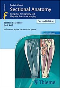
نام کتاب: Pocket Atlas Of Sectional Anatomy, Volume III – Spine, Extremities, Joints – Computed Tomography And Magnetic Resonance Imaging
نویسنده: Torsten Bert Möller و Emil Reif
ویرایش: ۲
سال انتشار: ۲۰۱۶
کد ISBN کتاب: ۳۱۳۱۴۳۱۷۲۵, ۹۷۸۳۱۳۱۴۳۱۷۲۱,
فرمت: PDF
تعداد صفحه: ۴۸۰
حجم کتاب: ۶۴ مگابایت
کیفیت کتاب: OCR
انتشارات: Thieme
Description About Book Pocket Atlas Of Sectional Anatomy, Volume III – Spine, Extremities, Joints – Computed Tomography And Magnetic Resonance Imaging From Amazon
Renowned for its superb illustrations and highly practical information, the third volume of this classic reference reflects the very latest in state-of-the-art imaging technology. Together with Volumes 1 and 2, this compact and portable book provides a highly specialized navigational tool for clinicians seeking to master the ability to recognize anatomical structures and accurately interpret CT and MR images.
Highlights of Volume 3:
New CT and MR images of the highest quality
Didactic organization using two-page units, with radiographs on one page and full-color illustrations on the next
Concise, easy-to-read labeling on all figures
Color-coded, schematic diagrams that indicate the level of each section
Sectional enlargements for detailed classification of the anatomical structure
Comprehensive, compact, and portable, this popular book is ideal for use in both the classroom and clinical setting.
درباره کتاب Pocket Atlas Of Sectional Anatomy, Volume III – Spine, Extremities, Joints – Computed Tomography And Magnetic Resonance Imaging ترجمه شده از گوگل
جلد سوم این مرجع کلاسیک که به دلیل تصاویر عالی و اطلاعات بسیار کاربردی مشهور است ، آخرین تکنولوژی تصویربرداری پیشرفته را منعکس می کند. این کتاب جمع و جور و قابل حمل ، همراه با جلد ۱ و ۲ ، یک ابزار ناوبری کاملاً تخصصی را برای پزشکان بالینی فراهم می کند که به دنبال تسلط بر توانایی تشخیص ساختارهای آناتومیک و تفسیر دقیق تصاویر CT و MR هستند.
نکات برجسته جلد ۳:
تصاویر جدید CT و MR با بالاترین کیفیت کتاب
سازمان تعلیمی با استفاده از واحدهای دو صفحه ای ، با رادیوگرافی در یک صفحه و تصاویر تمام رنگی در صفحه دیگر
برچسب زدن مختصر و آسان برای خواندن روی همه شکل ها
نمودارهای رنگی و شماتیک که سطح هر قسمت را نشان می دهد
بزرگنمایی های مقطعی برای طبقه بندی دقیق ساختار تشریحی
جامع ، جمع و جور و قابل حمل ، این کتاب محبوب برای استفاده در کلاس و کلینیک ایده آل است.
[box type=”info”]![]() جهت دسترسی به توضیحات این کتاب در Amazon اینجا کلیک کنید.
جهت دسترسی به توضیحات این کتاب در Amazon اینجا کلیک کنید.![]() در صورت خراب بودن لینک کتاب، در قسمت نظرات همین مطلب گزارش دهید.
در صورت خراب بودن لینک کتاب، در قسمت نظرات همین مطلب گزارش دهید.

