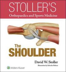
نام کتاب: Stoller’s Atlas Of Orthopaedics And Sports Medicine
نویسنده: David W. Stoller
ویرایش: ۱
سال انتشار: ۲۰۰۸
کد ISBN کتاب: ۹۷۸۰۷۸۱۷۸۳۸۹۷, ۰۷۸۱۷۸۳۸۹۵,
فرمت: EPUB
تعداد صفحه: ۱۰۳۹
حجم کتاب: ۱۲۵ مگابایت
کیفیت کتاب: اسکن
انتشارات: Lippincott Williams & Wilkins
Description About Book Stoller’s Atlas Of Orthopaedics And Sports Medicine From Amazon
Using 1,298 full-color anatomic drawings and 230 3-Tesla MR normal anatomy images, this atlas provides a detailed view of the intricacies of musculoskeletal anatomy. Dr. Stoller, through extensive cadaver dissections and imaging, has developed and proven new concepts on many musculoskeletal injuries, including hip impingement and patterns of meniscal tears. Muscles are shown in great detail, including origins and insertions. Skeletal structures are shown in relation to muscles, tendons, nerves, and ligaments. Clear legends describe the function and directional movement of muscles. Illustrations show both normal anatomy and mechanisms of injury, and Pearls and Pitfalls sections reinforce critical information.;MRI normal anatomy: lower extremity — MRI normal anatomy: upper extremity — The hip — The knee — The ankle and foot — Entrapment neuropathies of the lower extremity — Articular cartilage — The shoulder — The elbow — The wrist and hand — The finger — Entrapment neuropathies of the upper extremity — Marrow imaging — Bone and soft-tissue tumors.
درباره کتاب Stoller’s Atlas Of Orthopaedics And Sports Medicine ترجمه شده از گوگل
این اطلس با استفاده از ۱،۲۹۸ نقاشی آناتومیک تمام رنگ و ۲۳۰ تصویر آناتومی طبیعی ۳-تسلا MR ، نمای مفصلی از پیچیدگی های آناتومی اسکلتی عضلانی را ارائه می دهد. دکتر استولر ، از طریق تشریح و تصویربرداری گسترده جسد ، مفاهیم جدیدی را در مورد بسیاری از آسیب های اسکلتی عضلانی ، از جمله گرفتگی مفصل ران و الگوهای پارگی مینیسک ، ایجاد و اثبات کرده است. عضلات با جزئیات کامل ، از جمله ریشه و محل قرارگیری ، نشان داده می شوند. ساختارهای اسکلتی در ارتباط با عضلات ، تاندون ها ، اعصاب و رباط ها نشان داده شده است. افسانه های واضح عملکرد و حرکت ماهیچه ها را توصیف می کنند. تصاویر هم آناتومی طبیعی و هم مکانیسم های آسیب دیدگی را نشان می دهد و بخش های مروارید و دامها اطلاعات مهم را تقویت می کنند. نوروپاتی حبس اندام تحتانی – غضروف مفصل – شانه – آرنج – مچ دست و دست – انگشت – نوروپاتی محبوس در اندام فوقانی – تصویربرداری مغز – تومورهای استخوان و بافت نرم.
[box type=”info”]![]() جهت دسترسی به توضیحات این کتاب در Amazon اینجا کلیک کنید.
جهت دسترسی به توضیحات این کتاب در Amazon اینجا کلیک کنید.![]() در صورت خراب بودن لینک کتاب، در قسمت نظرات همین مطلب گزارش دهید.
در صورت خراب بودن لینک کتاب، در قسمت نظرات همین مطلب گزارش دهید.

